


Mammography
Mammography - What is it?
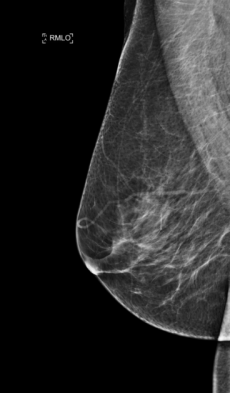
Mammography is a special X-ray examination of the mammary gland. The soft tissue of the breast hardly differs in density, so soft X-ray beams are used for imaging to obtain a high-contrast image. For the examination, the breast is placed between the X-ray tube and the film table and carefully compressed to a thickness of approximately 4-6 cm for a few seconds using a Plexiglas cylinder. This procedure is often described by patients as unpleasant, but it is unavoidable. The compression avoids inaccuracies due to movements and the tissue of the breast is evenly distributed, thus the image becomes even clearer. Furthermore, this makes the radiation exposure for the patient even less than it already is. For each breast, 2 images are taken, with the radiation path running from top to bottom or from the inside obliquely down to the outside at the top. Only mammography can detect microcalcifications, which can be an expression of breast cancer or breast precancer. Additional special images (e.g. magnification images) and tomosyntheses (slice images) are possible for more precise assessment.
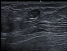
In addition, sonography of the breast is performed if the breast tissue is so dense that mammography can only be assessed to a limited extent. Sonography can be used to differentiate between benign fluid-filled cysts and malignant tumors.
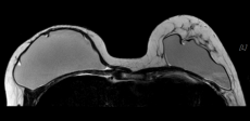
In special cases, an MRI of the breast is necessary, e.g., to clarify unclear findings in mammography and sonography, to differentiate between scar tissue and tumor recurrence after breast cancer surgery, or to evaluate breast implants.
At our facility, specimen collection can be performed under mammographic, sonographic, or MR tomographic view. Wire markings of tumor foci can also be performed in this manner prior to surgery.
Galactographies are performed to visualize tumors in the milk ducts. This involves carefully probing a secreting milk duct, injecting a contrast agent, and then obtaining a mammogram.
Mammography - For whom?
Mammography provides us with a diagnostic procedure that is primarily used for screening and early diagnosis of tumors of the breast. According to general recommendations, the first preventive mammography should be performed around the age of 35. From the age of 40 to 70, a follow-up examination is then recommended at regular intervals of 2 years. If there are risk factors that promote the occurrence of breast cancer (previous breast cancer, breast cancer of the mother or sister, hormone therapy during menopause), the checks should be performed annually. This screening procedure makes it possible to detect tumors at a very early stage.
Mammograms are performed to clarify symptoms such as palpable lumps, skin or nipple retractions, pain, inflammation, or secretion. Mammograms are used to take tissue samples from the breast and wire-mark tumor sites. Patients with breast cancer receive mammograms for treatment planning and progress monitoring, as well as during follow-up care.